A radiologist looking at a brain scan sees texture, shape, edges. A radiomics pipeline sees hundreds of numbers extracted from those same pixels, fed into a machine learning model that returns a prediction. Somewhere between the two sits a gap that has kept most of this research off hospital workstations: the clinician cannot see what happened in the middle.
A team led by Yin Lin, working with colleagues at institutions in Italy including a neuro-oncology group, has released the first version of a web-based system built to close that gap. Their paper, posted to the arXiv preprint server on 29 July 2026, describes a single browser interface that handles three jobs which normally live in three separate places: organizing a patient cohort from a clinical spreadsheet, pulling radiomic features out of medical images and their segmentation masks, and running those features through pre-trained machine learning models.
The word that does the most work in their description is "guarded." Inference in this system is not a button that always returns an answer. The authors describe guarded inference with pre-trained models, meaning the platform checks conditions before it will produce a prediction, rather than accepting whatever a user uploads and returning a number regardless.
What the system actually exposes
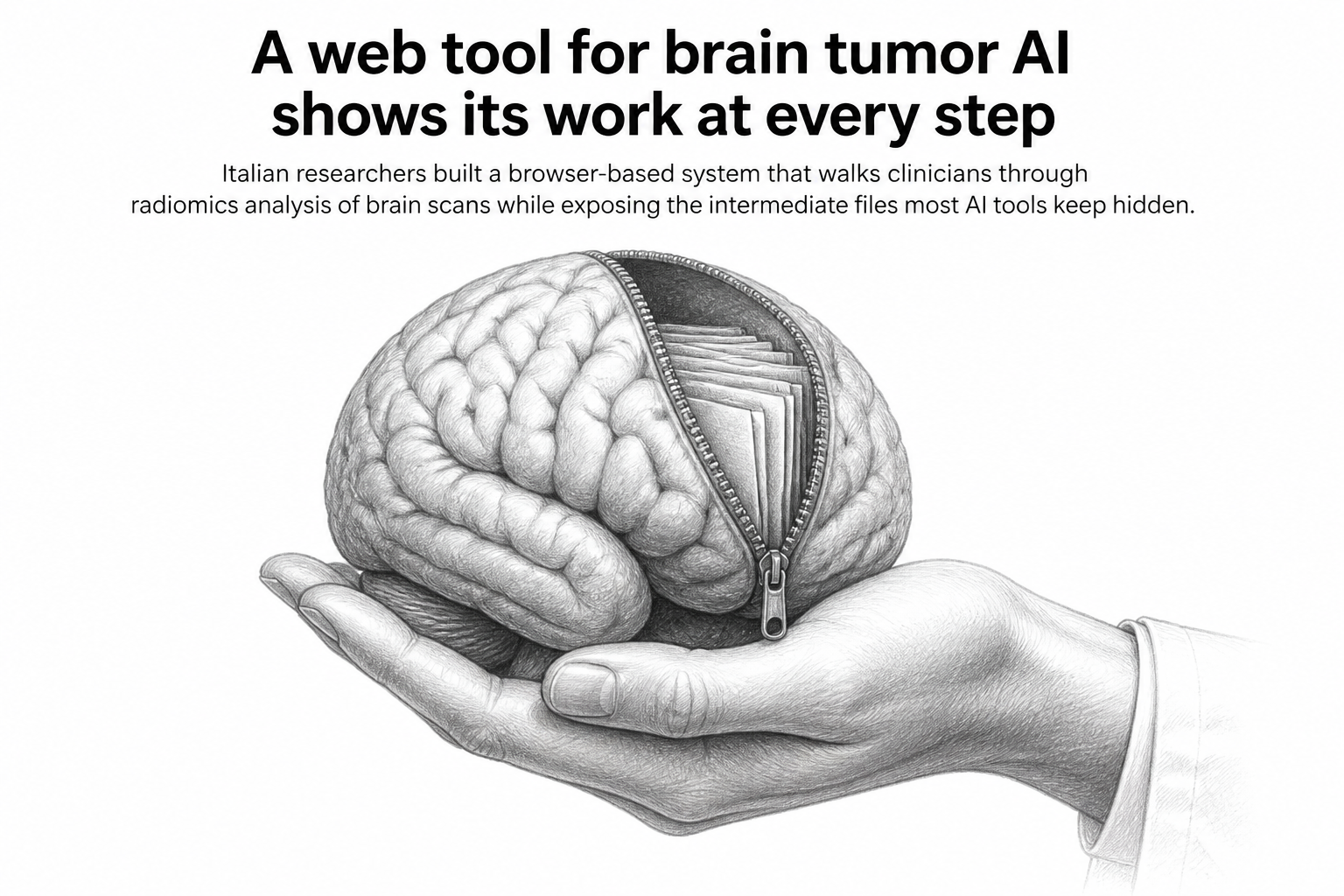
The contribution the authors single out is not the accuracy of any model. It is the explicit exposure of intermediate workflow artifacts. In an ordinary radiomics pipeline, a scan goes in and a risk score comes out, with the feature tables, the masks, and the intermediate transformations living in scripts on somebody's laptop. This platform surfaces those artifacts in the interface. A user can look at what the system extracted, in what order, and on which images, before deciding whether to trust the model's output.
That design choice sounds modest. It addresses something the authors name directly as one of three barriers holding radiomics back from clinical use: fragmented workflows, poor transparency, and weak integration with what end users actually need. The first and third of those are as much software engineering problems as scientific ones, which is likely why the paper was filed under software engineering rather than machine learning or medical imaging.
The team did not design the system in isolation and hand it over. They describe an iterative user-centred design process, meaning the intended clinical users shaped the interface as it was being built. They then evaluated the platform on two datasets: a public glioblastoma dataset, and a proprietary clinical cohort. Glioblastoma is the most aggressive common primary brain tumor in adults, and it is the disease radiomics research returns to most often.
What the paper does not claim
Here the reporting has to be careful, because the paper itself is careful. This is a preprint, which means it has not yet been through peer review. Only the abstract and metadata are publicly available in the record consulted for this article, so the specific evaluation results, the number of patients in either cohort, and the performance of the pre-trained models are not stated in the material at hand.
The authors do not claim the system improves diagnosis. They do not claim it is ready for clinical deployment. What they claim is narrower and more honest: that combining portability, inspectability, and simple deployment gives a practical foundation for clinically oriented AI in brain tumor analysis. A foundation is not a finished building. They call this the first version, and they mean it.
Why it matters
Most of the difficulty in getting medical AI into hospitals is not that the models are bad. It is that a research pipeline and a clinical tool are different objects. A research pipeline can be a folder of Python scripts run by the person who wrote them. A clinical tool has to run on someone else's machine, be operated by someone who did not build it, and produce output that a clinician can defend to a colleague or a patient.
The emphasis on traceability speaks to a specific professional anxiety. A neuro-oncologist asked to act on a model's output needs to know where that output came from. If the answer is a black box, the reasonable response is to ignore it. By putting the intermediate steps on screen, the authors are betting that inspectability is what earns a tool a place in the workflow, more than a marginal gain in accuracy would.
There is a broader point in the choice of venue and framing. Filing this under software engineering is an argument in itself: that the bottleneck in clinical AI is increasingly about deployment, interfaces, and trust rather than model architecture. Whether this particular platform gets adopted is an open question, and the evidence in the abstract is not enough to answer it. The problem it identifies, however, is one that a great deal of well-performing medical AI research has run into and failed to get past.