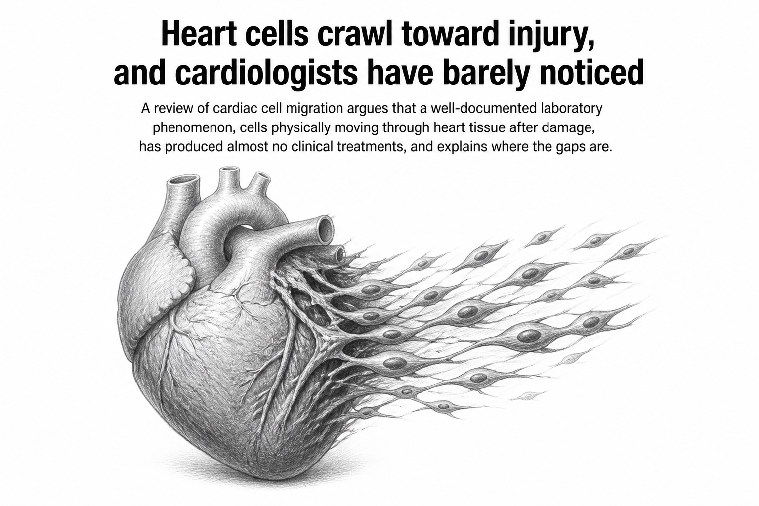
Two days after a piece of heart muscle is cut away in a lab animal, the muscle cells left behind start to move. They push out projections and creep toward the wound, and they do it hugging the new blood vessels sprouting into the damaged zone. Sixty-nine percent of those projections sit within 15 micrometres of the nearest vessel, about the width of two cell nuclei. The average gap is 12.4 micrometres.
That tight spacing is a clue, and it is one of many collected in a new review by Jie Ma, Yaping Xu, Yali Wang and Zhikun Guo of Henan Medical University and Zhengzhou Seventh People's Hospital, published in the International Journal of Medical Sciences. Their argument is in the title: cardiac cell migration is a busy research field that has not attracted clinical attention. Thousands of experiments describe cells crawling around inside injured hearts. Almost none of that has turned into a treatment.
Why do migrating heart muscle cells stay so close to new vessels? The authors point to a shared chemical signal. In zebrafish with amputated ventricles, a molecule called SDF-1α, released by the heart's outer layer, pulls muscle cells toward the injury. Its receptor, CXCR4b, sits on muscle cells but not on the vessel-lining endothelial cells. Block the receptor and the muscle cells stall while the vessels carry on regardless. Because a closely related receptor appears on both cell types, the team suggests one chemical gradient may be steering vessel growth and muscle-cell movement at once.
The traffic report after a heart attack
Most of the review is a timetable of who arrives when. Within five minutes of a heart attack in mice, macrophages, the immune system's cleanup cells, are already piling into the dead zone. Circulating monocytes begin crossing into injured muscle by thirty minutes. Within a day, the heart's original resident macrophages are nearly gone, replaced by newcomers from bone marrow and spleen; the spleen alone supplies up to half of them, mobilised by the blood-pressure hormone angiotensin II. Around ten days in, fibroblasts move through to build scar tissue. Endothelial cells and pericytes, the cells that wrap around capillaries, arrive to rebuild the blood supply.
Sequence matters. When researchers removed the tip of the heart in newborn mice, endothelial cells reached the wound first and grew into working arteries, and only then did the muscle cells migrate in. The plumbing goes in before the tenants.
Almost every cell in this story cuts both ways. Neutrophils clear dead tissue early, but if too many arrive, or they linger, they flood the area with reactive oxygen species and inflammatory signals that cause a second wave of damage. Pericytes secrete factors that build new microvessels, then, under the influence of the growth factor TGF-β1, some convert into myofibroblasts that lay down collagen and stiffen the heart. SDF-1α, the navigation beacon, also recruits so many inflammatory macrophages that the electrical coupling between muscle cells goes haywire, slowing conduction across the ventricle. The authors describe the ideal intervention as selectively recruiting stem cells while blocking the inflammation that rides along with them. Nobody has managed it.
The same cast behaves differently in chronic heart failure. There, the migration never resolves. Neutrophils trickle in continuously instead of surging and clearing. Macrophages stay stuck in their inflammatory M1 state rather than switching to the reparative M2 form. Fibroblasts keep migrating and keep depositing matrix until the muscle is stiff. Endothelial cells lose their ability to build vessels, and capillaries disappear. Regulatory T cells, protective after a heart attack, can convert into inflammatory Th17 cells that drive fibrosis instead.
The authors are frank about why so little has translated. Most migration work happens on rigid two-dimensional plastic or in static collagen gels, neither of which resembles living heart tissue with its viscoelasticity, shifting stiffness, and uneven fibres. Cells in those models move in different modes and at different speeds than they do in a body. Live imaging inside animals has poor resolution and weak signal and cannot track cells for long. And the drug targets people have identified, molecules like Rho and focal adhesion kinase, are broad-spectrum machinery used by nearly every moving cell, so blocking them has heavy side effects.
The review's hope rests on spatial omics: techniques that read gene or protein activity while keeping each cell's position in the tissue intact. Conventional single-cell sequencing dissolves the tissue first, so you learn what a migrating cell is but not where it came from or where it was headed. Methods such as MERFISH and seqFISH keep the map.
Why it matters
Heart attacks and heart failure are, at bottom, problems of repair. The muscle that dies is mostly replaced by scar, and adult human heart muscle renews itself only slightly each year, largely through existing muscle cells dividing rather than through progenitor cells, a point on which the field's consensus shifted after early claims failed to replicate in independent labs.
This review does not report a new experiment or a new drug. It is a stocktaking, and its value is in naming the gap plainly: the biology of cardiac cell migration is documented in fine detail, and the clinical pipeline built on it is close to empty. The authors suggest a workable near-term goal, drugs that block the pathological migration driving fibrosis and runaway inflammation without disturbing the physiological kind. Whether the signals can be separated that cleanly is exactly what remains unknown. Note too that nearly all the evidence here comes from mice, rats, and zebrafish; the authors repeatedly flag how little is known about whether adult human heart muscle cells migrate at all.