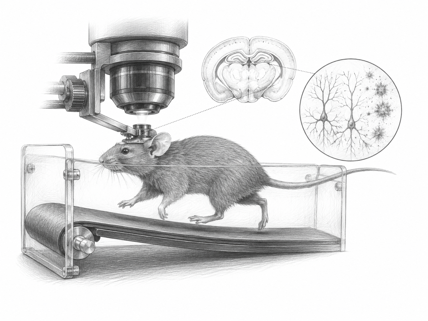
A mouse runs on a treadmill under a microscope, and in the memory-forming part of its brain, thousands of neurons should be doing two things at once: staying relatively quiet while the animal is still, then firing hard when it moves. In mice carrying five human Alzheimer's mutations, both halves of that pattern went wrong. The quiet got noisier. The firing got weaker. What was left was a narrower band between rest and motion, less room, in effect, for the brain to say anything at all.
That compression is the central finding of a new preprint from Mary Ann Go, Simon Schultz and colleagues at Imperial College London, with collaborators in Melbourne and Manila. The team used two-photon calcium imaging, a technique that watches individual neurons light up in a living, behaving animal by tracking the calcium that floods in when a cell fires, to record hippocampal CA1 activity in 5xFAD mice, a widely used strain engineered to build up amyloid plaques early and aggressively.
CA1 is a reasonable place to look. It is a thin output layer of the hippocampus, and it is where researchers first found place cells: neurons that fire when an animal occupies one particular spot, and together form something like an internal map. If amyloid degrades memory by degrading circuits rather than just killing cells, CA1 is where the damage should be legible.
What compressed range means
The authors report elevated baseline activity in the 5xFAD animals alongside reduced locomotion-driven firing. Those two abnormalities pull in opposite directions but add up to the same problem, which the authors call a diminished dynamic range, the gap between a neuron's floor and its ceiling. A dial that only turns from four to six cannot carry as much information as one that turns from one to ten. To the authors' knowledge, this is the first direct experimental evidence for reduced dynamic range in an Alzheimer's model, a prediction that has circulated in the field without measurement behind it.
Crucially, the effect was not uniform across the tissue. The abnormalities were strongest in neurons near amyloid plaques, and they spread more widely as the animals aged. That spatial gradient is the part of the result that does real explanatory work: it ties a local, physical piece of pathology to a specific failure in how nearby neurons behave, rather than leaving "amyloid" and "dysfunction" as two facts sitting side by side.
The team also saw altered synchrony across the network, changes in how tightly neurons fired together, along with degraded spatial coding and more variable responses from cell to cell and trial to trial. Variability matters more than it sounds. A neuron that gives a slightly different answer each time the mouse passes the same landmark is a neuron whose downstream partners cannot trust it.
Then there is timing. When the researchers watched place fields form, they emerged more slowly in the 5xFAD mice than in controls, and this held in both familiar and novel environments. The authors read the familiar-environment delay as impaired recall and the novel-environment delay as impaired learning. Two different cognitive jobs, one shared bottleneck.
Why it matters
Most of what is known about how Alzheimer's disrupts the brain comes from the two ends of the problem: molecules and plaques at one end, behaviour and clinical decline at the other. The middle, what circuits of neurons actually do differently while an animal is awake and navigating, is harder to reach and correspondingly thinner. Work like this fills in that middle, and the specific claim here, that pathology narrows the range over which neurons can be flexibly modulated, is a different kind of explanation than cell death. A compressed dial is still a working dial. That leaves at least the conceptual possibility of restoring range, which a dead neuron does not offer.
The caveats are real and worth holding onto. These are mice, and 5xFAD mice in particular: they carry five mutations at once and accumulate amyloid far faster and more floridly than any human does, which makes them useful for seeing amyloid's effects clearly and unreliable as a model of the human disease's pace. The paper is a preprint, posted to bioRxiv on 28 July 2026 and not yet through peer review, though its authors note this version was substantially revised, reanalysed and rewritten from the original. The abstract reports directions of effect, elevated, reduced, more variable, without the effect sizes that would let a reader judge magnitude, so how badly the range compressed is not something to state from what is public here.
What the study offers is a mechanism specific enough to argue with. If amyloid plaques locally squeeze the operating range of the neurons around them, and if that squeezing spreads with age, then the slow arrival of a place field in a mouse and the slow arrival of a memory in a person may turn out to be versions of the same failure. That is a hypothesis, not a result. It is the kind worth testing.