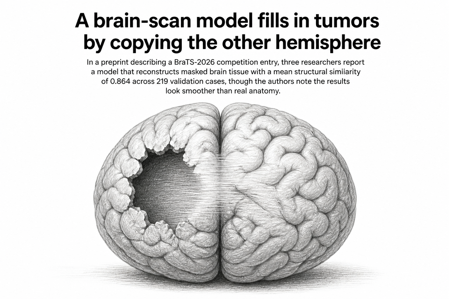
Cut a hole in a T1-weighted brain MRI and ask a computer to fill it back in with plausible healthy tissue. That is the whole of the ASNR-MICCAI BraTS Local Synthesis task, and it is less strange than it sounds. Tumors distort the anatomy around them, which makes many downstream measurements awkward: you want a picture of what the brain looked like before the lesion arrived. Inpainting is one way to get a tumor-free reference to compare against.
Danilo Danese, Angela Lombardi and Tommaso Di Noia, writing in a preprint posted to arXiv on 30 July 2026, describe their entry to that competition. Their reported score on the official BraTS-2026 validation leaderboard: a mean structural similarity of 0.864 in the healthy region, a peak signal-to-noise ratio of 24.7 decibels, and a mean squared error of 4.6 thousandths, measured over 219 cases.
The design choices behind those numbers are the interesting part. The competition scores submissions on distortion metrics, which measure how close the filled-in pixels are to the ones that were removed. So the team built a deterministic regression model, one that produces a single best guess rather than sampling from a range of possible completions, and then spent their effort on giving that model the right instincts for the job.
Two ideas about where to look
The network is a volumetric encoder-decoder, meaning it processes the scan as a three-dimensional block rather than slice by slice, and it follows what the authors call the U-DiT principle: run self-attention on a downsampled grid of tokens. Self-attention is the mechanism that lets a model relate distant parts of an image to each other, and it is expensive, so shrinking the grid first keeps the cost manageable. Convolutions and skip connections, which pass fine detail directly from the encoder to the decoder, hold on to the sharp edges that the downsampling would otherwise blur away. Three-dimensional rotary position embeddings tell the attention block where in the volume each token sits.
The first of the team's two central ideas is a constraint on that attention. Tokens inside the masked hole, which the authors call "void" tokens, are allowed to attend only to tokens known to be healthy in the same scan. They cannot look at each other. The reasoning is direct: if unknown regions can inform other unknown regions, the model can talk itself into a self-consistent fiction. Forcing every void token to draw from observed anatomy keeps the completion anchored to something real. On top of that, the model learns a bias steering each query toward its contralateral homologue, which is the matching structure on the opposite side of the brain.
The second idea is the same intuition, applied more bluntly. The team feeds the model a mirrored copy of the scan as an extra input, so the healthy hemisphere is handed over directly as a patient-specific template. Brains are roughly bilaterally symmetric and tumors usually appear on one side, so the intact half is often a decent stand-in for the missing half of the same patient. The authors report that this prior improves the distortion metrics at matched structural similarity.
The smoothness problem
The paper does not stop at the leaderboard number. The authors also analyse what they call the residual smoothness inherent to distortion-optimal regression, and discuss what it means for anatomical realism. The point is worth sitting with. When a model is trained to minimise average error and there are many plausible ways to fill a hole, the safest output is something like the average of all of them. Averages are smooth. A blurred compromise between several possible cortical folding patterns will score well on pixel-wise distortion metrics while looking, to a radiologist, subtly unlike a brain.
That is a tension the authors raise themselves rather than one imposed from outside, and it is a limit on what the score means.
Why it matters
Synthetic healthy tissue is not a diagnosis and nobody here suggests otherwise. Its use is more mundane and more practical: giving analysis pipelines a version of the scan without a lesion in it, so that tools built to measure normal anatomy have something to work with.
The attention constraint is the idea most likely to travel. Telling a model which parts of its input it is not allowed to consult, and encoding an anatomical fact (bilateral symmetry) as a structural rule rather than hoping the network infers it from data, is a general move. It applies wherever a domain has known structure and training data is scarce, which describes most of medical imaging.
Two caveats sit on top of all of it. This is a preprint on arXiv, not yet peer-reviewed, and the numbers come from a competition validation leaderboard rather than from any clinical evaluation. The available text is the abstract page; the fuller methods and comparisons live in the PDF. And the authors' own closing observation deserves the last word: a model can win on distortion and still produce tissue that does not quite look alive.