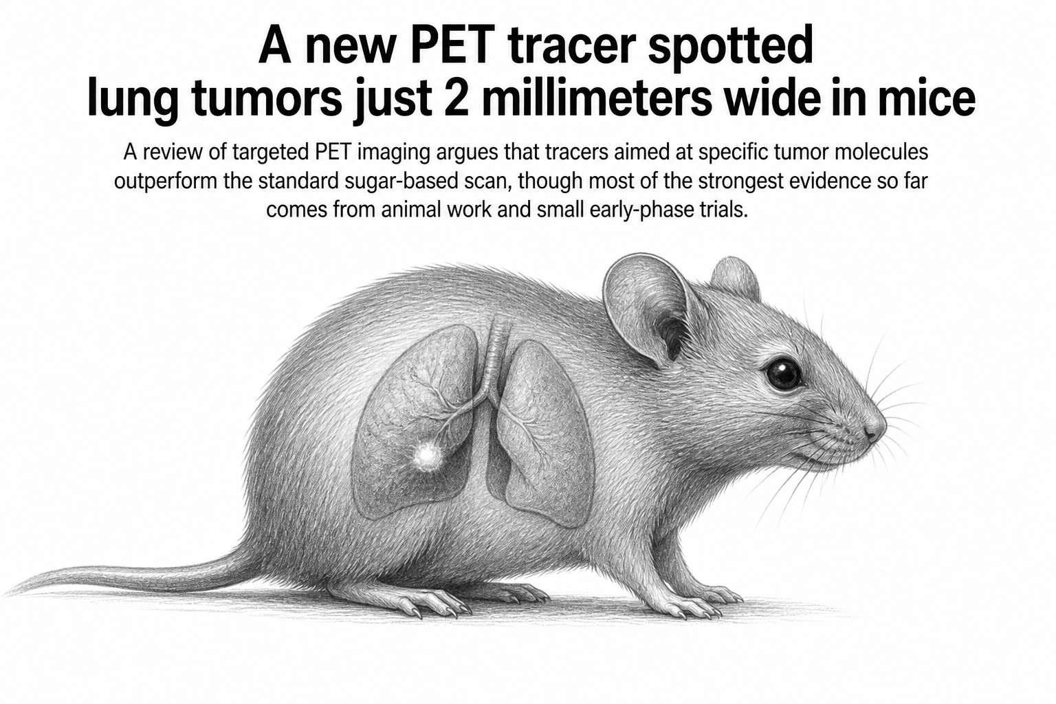
Nine point nine three. That is the tumor-to-background ratio a team at the Jiangsu Institute of Nuclear Medicine measured when they imaged mice carrying tiny malignant lung nodules with a radioactive antibody aimed at a protein called CEACAM6. The standard clinical scan, fluorodeoxyglucose PET, managed 2.68 in the same animals. The nodules averaged 1.39 millimeters across, roughly the thickness of a credit card, and the targeted probe found them.
That contrast is the argument running through a new review in Chemical & Biomedical Imaging by Chongyang Chen, Min Yang and colleagues, who survey where molecular PET imaging for lung cancer currently stands. Their case starts with a problem clinicians know well. Low-dose CT scanning catches lung cancers early and has measurably cut deaths from the disease, but it sees shape, not biology. It cannot reliably tell a malignant nodule from a scar, an old infection, or a patch of inflammation. False positives run at 20 to 40 percent, the authors write, which means a great many people undergo follow-up procedures they never needed.
PET was supposed to fill that gap. The workhorse tracer, 18F-FDG, is a radioactive sugar analogue: cancer cells tend to burn glucose fast, so they light up. The trouble is that inflamed tissue burns glucose fast too, and some lung adenocarcinomas barely burn it at all. The scan measures metabolism, not malignancy, so it produces both false alarms and missed tumors.
Aiming at the tumor instead of its appetite
The alternative is to build tracers that bind something specific to the cancer. Attach a positron-emitting isotope to an antibody, a small peptide, or a drug-like molecule that recognizes a particular protein, inject it, and the scanner maps wherever that protein lives in the entire body.
The review catalogues a long list of these targets. Some are the oncogenic drivers that already guide treatment, such as EGFR and MET. Others sit in the tissue surrounding the tumor. Fibroblast activation protein, or FAP, marks the cancer-associated fibroblasts that build a tumor's supportive scaffolding, and FAP tracers have produced some of the most striking clinical numbers here. In one head-to-head comparison of 34 patients with advanced lung cancer, 68Ga-FAPI PET/CT flagged more suspected metastases than FDG at every site the authors list: 356 versus 320 lymph nodes, 109 versus 91 bone lesions, 66 versus 35 in the pleura, and 23 versus 10 in the brain. A pooled sensitivity figure for detecting metastatic lesions came out at 99 percent for FAPI against 77 percent for FDG.
Immune imaging is the other active front. PD-L1, the protein that immunotherapy drugs are designed around, is currently assessed by staining a biopsy sample, which captures one spot in one lesion at one moment. A whole-body scan does not have that limitation. In a prospective study of operable non-small-cell lung cancer patients receiving chemotherapy plus immunotherapy before surgery, baseline uptake of the PD-L1 tracer 68Ga-NOTA-WL12 correlated strongly with which patients had a major pathological response. FDG PET, by contrast, mostly tracked whether tumors shrank and predicted nothing.
What the numbers do not settle
The authors are careful about how far any of this has travelled. Much of the most dramatic data, including the CEACAM6 result and a companion probe targeting the checkpoint protein FGL1, comes from mice. The clinical studies that exist are largely first-in-human safety work or single-centre series with dozens of patients, not the thousands that change guidelines.
And there are structural obstacles that no amount of tracer chemistry fixes. Target proteins vary from one region of a tumor to another and from one metastasis to the next, so a single scan at a single moment can mislead. Expression also shifts during treatment, sometimes faster than the follow-up interval. Building the tracers is its own bottleneck: manufacturing must meet pharmaceutical-grade standards, short-lived isotopes like carbon-11 require a cyclotron on site, and antibody-based probes labelled with zirconium-89 need hours or days between injection and scan, raising the radiation dose. Regulators and insurers, the authors note, are set up to evaluate tests that produce a yes or no decision, not ones that add texture to a picture already assembled from biopsies and blood tests.
Why it matters
Lung cancer kills more people than any other cancer, and most of that toll traces back to two failures: finding tumors too late, and giving the wrong patients the wrong drugs. Both are information problems.
A scan that reports which molecules a tumor is actually displaying, across every lesion in the body at once, addresses something a needle biopsy structurally cannot. Whether that translates into people living longer is the question the field has not yet answered, and the authors say so plainly. What they document instead is a set of tools that measure the right things, waiting on the large prospective trials that would show those measurements change what doctors do.
One detail hints at where this goes next. The same molecular target that makes a good imaging beacon can carry a therapeutic isotope instead of a diagnostic one. The CEACAM6 antibody labelled with zirconium-89 finds tumors; the same antibody labelled with iodine-131 shrank them in mice. That pairing already works in prostate cancer and neuroendocrine tumors. Lung cancer is the harder case, and it is next in line.