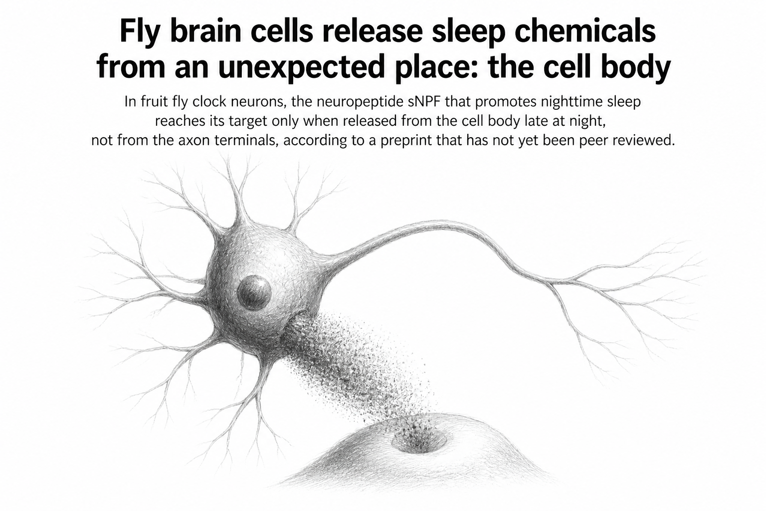
The textbook picture of a neuron talking to its neighbor is tidy. A signal travels down a long axon, arrives at the terminal, and chemicals spill across a tiny gap into the next cell. In the fruit fly brain, two sets of clock neurons appear to break that rule entirely.
The cells in question are the small and large ventral lateral neurons, known as s-LNv and l-LNv. Together they help set the fly's daily rhythms and its sleep. Earlier work established that s-LNv neurons promote nighttime sleep by releasing a short peptide called sNPF, which acts on receptors sitting on l-LNv neurons. The awkward detail is that the two cell types are not wired to each other by chemical synapses at all. So how does the message get across?
A team led by Markus Klose and Edwin Levitan, working across the University of Pittsburgh, Johns Hopkins University Applied Physics Laboratory, Peking University, Carnegie Mellon University and Cornell University, went looking for the answer by watching sNPF move through the adult fly brain directly. Rather than inferring the peptide's route from behavior, they imaged where it actually spread.
What they saw did not match the obvious guess. The s-LNv neurons do produce a burst of sNPF from their axon terminals every day in the middle of the morning, a well-known event. But that burst, the team reports, never reaches the l-LNv neurons. It does not even reach the s-LNv cell bodies, the rounded hubs of the neurons themselves. The morning release, in other words, goes somewhere else.
The signal that does arrive comes from a different place and a different time. Late at night, the s-LNv cell body itself releases sNPF, prompted by IP3 signaling, an internal chemical cascade the authors link to promoting sleep. That peptide reaches the cell bodies of l-LNv neurons. It does not reach their terminals. Two releases of the same molecule from the same neuron, hours apart, and each one lands in a place the other cannot.
This matters because release from the cell body is not how neurons are usually described as communicating. Cell bodies are typically cast as the metabolic headquarters, not the broadcast antenna. Here the soma appears to be doing the behaviorally important work, at least for this particular sleep signal.
Threads between neurons
Diffusing peptides were not the whole story. The team also examined fly connectomes, the painstaking wiring maps assembled from electron microscopy of the fly brain, and found that adult s-LNv and l-LNv neurons touch each other directly through cytonemes. Cytonemes are thin cellular projections that reach out and make contact without forming a chemical synapse. The connection is physical but not synaptic in the classical sense.
When the researchers genetically disrupted these cytonemes in LNv neurons, the effect was oddly specific. Sleep latency changed, meaning the time it takes a fly to fall asleep. Nighttime sleep itself did not change, which is the behavior driven by the somatic sNPF release. Neither did circadian behavior, the fly's underlying daily rhythm, which depends on a different peptide called PDF released from the terminals.
That separation is the paper's central claim. Three parts of the same neurons, the terminals, the cell body, and the cytonemes, each control a different slice of rhythmic behavior, and interfering with one leaves the others intact. It is an unusually clean division of labor inside a single cell.
The authors add a note of caution about the cytoneme contacts. These contacts, they write, are possibly acting through connections that current connectome annotations do not capture. The wiring diagrams that neuroscientists rely on catalogue chemical synapses; a contact that is not a synapse can sit in the data unlabeled.
Why it matters
Connectomes have become one of the main tools for reasoning about how brains produce behavior. The premise is that if you map every synapse, you can in principle trace how a signal moves. This work suggests the map has blind spots that matter. A neuron can release a peptide from its cell body that travels to a target it has no synaptic connection with, and it can touch that target through projections the annotation scheme does not record.
The practical caution is worth stating plainly. If two neurons show no synaptic link in a connectome, that is not the same as showing they do not communicate. Anyone reading a wiring diagram as a complete account of a circuit may be missing routes like the ones described here.
Some limits are worth keeping in view. This is a preprint posted to bioRxiv on July 30, 2026, so it has not been through peer review. The work is entirely in Drosophila, and fly clock neurons are not human sleep circuitry. Whether release from the cell body plays a comparable role in larger brains is not something this paper tested or claims.
What it does offer is a concrete demonstration that a neuron's cell body is not merely support infrastructure. In these flies, it is a sender in its own right, on its own schedule, reaching a target its own axon terminals cannot.