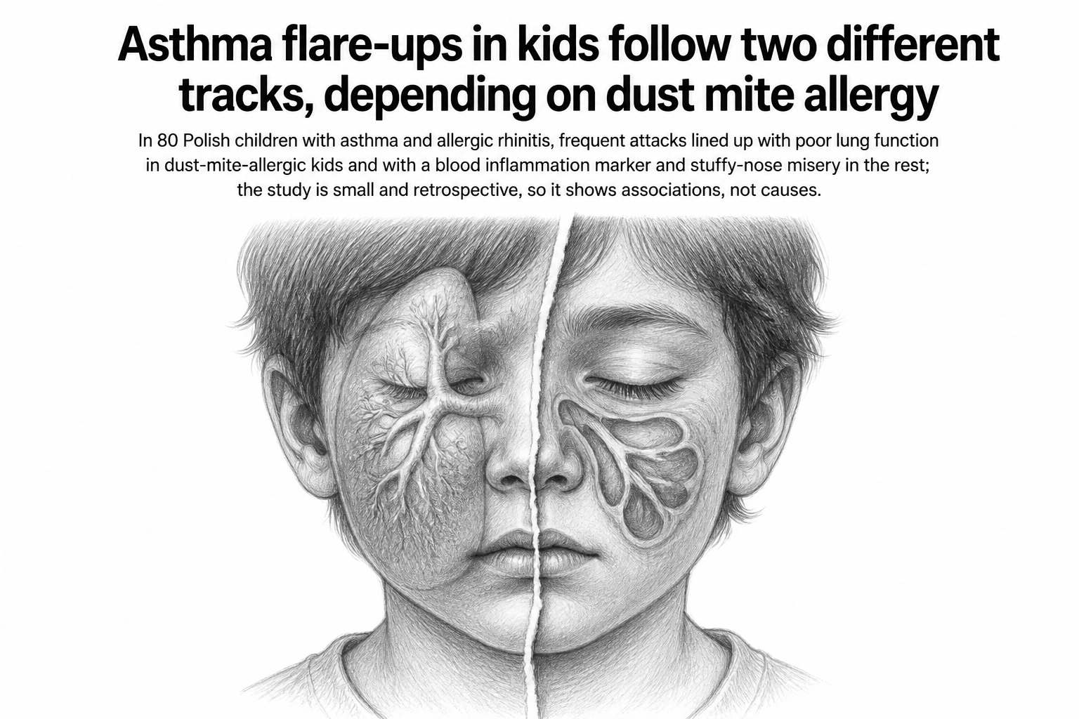
Thirty-eight of the children had gotten through the past year with at most one asthma attack. Forty-two had two or more: the kind of acute worsening that means a course of steroid pills, an unplanned clinic visit, a trip to the emergency department, or a hospital bed. All 80 were patients at the Medical University of Lodz in Poland, aged 4 to 18, all with both asthma and allergic rhinitis. When Katarzyna Gmachowska and colleagues looked for what separated the frequent flare-ups from the rare ones, the answer turned out to depend on which child they were looking at.
The team split the group by a single allergy: sensitization to house dust mite, confirmed from earlier skin prick tests or blood antibody levels. Forty-four children were sensitized, thirty-six were not. Then they ran the same battery on everyone during a period of clinical stability, after any acute symptoms had settled. Spirometry for lung function. Exhaled nitric oxide, a breath test that tracks a particular flavor of airway inflammation. Blood eosinophil counts. A skin-autofluorescence reading used as an indirect stand-in for activity in the AGE-RAGE pathway, an inflammation and tissue-damage signaling system that has drawn interest across lung diseases. Two questionnaires: one scoring asthma control, one scoring how much sinus and nasal symptoms drag on daily life.
Among the dust-mite-allergic children, the frequent-flare group breathed measurably worse. Their median FEV1, the volume of air forced out in one second as a percentage of predicted, sat at 87 percent against 98 percent in the infrequent group (p = 0.0089). The FEV1/FVC ratio, a standard index of airflow obstruction, was lower too (p = 0.0186). Their asthma control scores were worse (p = 0.0117). It is the picture a pulmonologist would expect: narrowed airways, poorly controlled disease, repeated attacks.
The children without dust mite allergy showed none of that. Lung function was statistically indistinguishable between their frequent and infrequent flare groups. What did separate them was sRAGE, the soluble decoy form of the RAGE receptor, which was higher in the children with more attacks (p = 0.0259), and how much their nose and sinus symptoms limited daily activity (p = 0.0309). Same disease label, same clinic, different fingerprint.
The breath test that stayed quiet
One result is notable for what it failed to show. Exhaled nitric oxide and blood eosinophil counts, the two workhorse markers of type 2 inflammation, did not differ between frequent and infrequent flare-ups in either subgroup. Not in the allergic children, not in the others. The authors are careful about why: nitric oxide readings are pushed down by inhaled steroids, and these children stayed on their normal treatment throughout. Blood eosinophils came from a single lab draw pulled retrospectively from medical records, which the authors flag as a limitation, since eosinophil counts wander over time.
A separate look across all 80 children tied the nose to the chest. Among those with milder sinonasal symptoms, 78 percent had good asthma control. Among those with more severe symptoms, only 45 percent did (p = 0.012). Put the other way, poor control was roughly two and a half times as common in the stuffier group. The nasal lining and the bronchial lining sit on a continuum, anatomically and immunologically, and the authors read this as inflammation upstairs feeding instability downstairs.
Why it matters
Asthma is not one disease, and the treatments increasingly are not one treatment either. Sorting children by which biological process is actually driving their attacks is the practical goal, and the standard sorting tools here did nothing. What did discriminate was cheap and already sitting in the clinic: a spirometer for the dust-mite-allergic kids, and a five-question survey about a blocked, itchy nose for the rest.
The sRAGE finding is the more speculative half. Earlier work has pointed in opposite directions, with some studies finding sRAGE lower in asthmatic children and reading it as protective. Here it was higher in the children flaring most often. The authors suggest this might be the body ramping up production in response to ongoing inflammation, but they present it as a hypothesis and say the RAGE axis in children's airways needs mechanistic work before anyone reads much into a number.
The honest limits are considerable, and the paper states them plainly. Eighty children is small. The design is cross-sectional with attack history collected retrospectively, so lung function was measured after the flare-ups had already happened, which means nobody can say which came first. Biomarkers were single snapshots. No multivariable analysis was run, so age, sex, treatment intensity, and underlying severity remain unadjusted. Larger prospective cohorts are the stated next step.
What survives all that is a usable suggestion. When a child's asthma keeps flaring and the standard inflammation numbers look unremarkable, the answer may be sitting one floor up, in the nose.