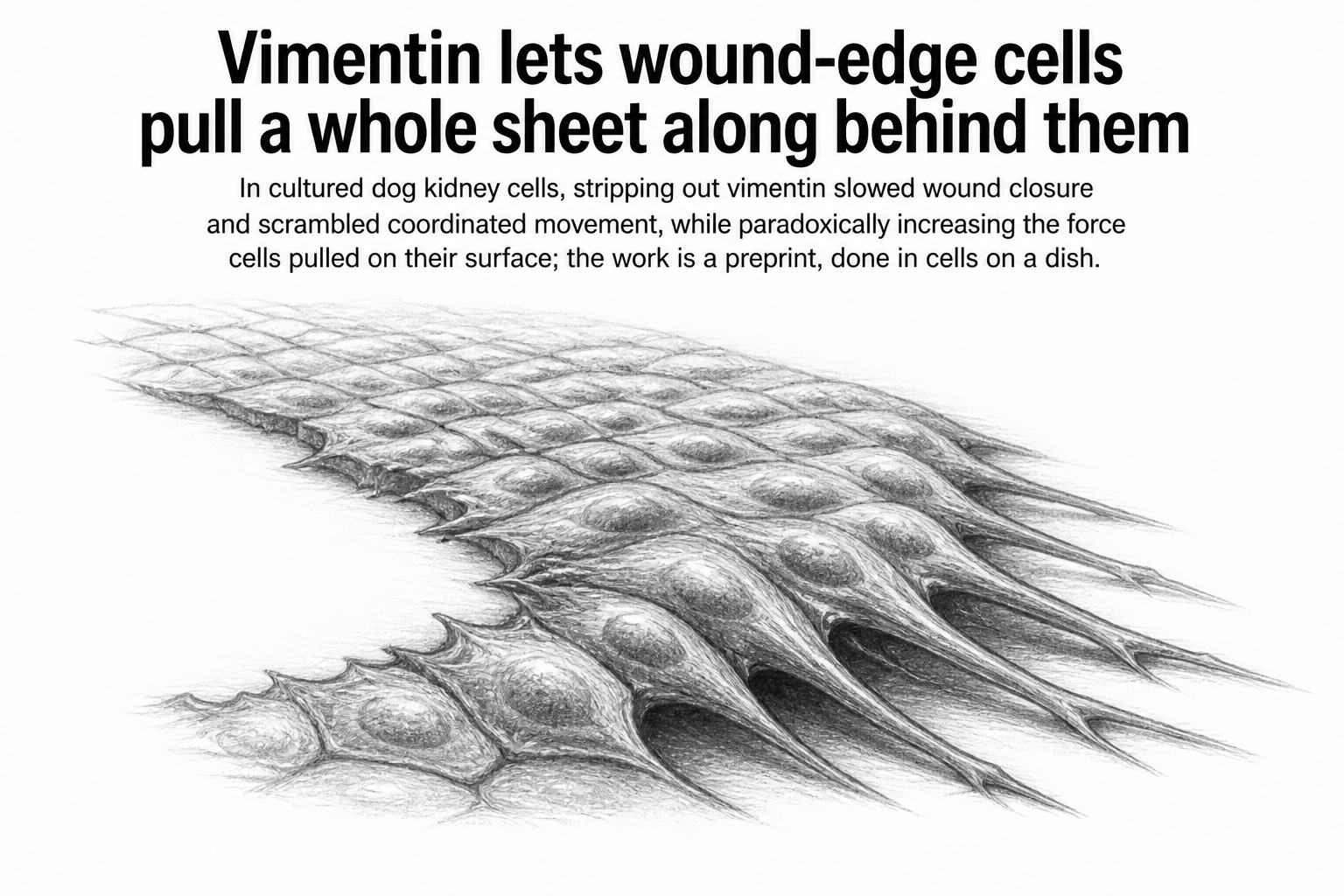
Scratch a sheet of cells growing in a dish and the cells at the torn edge change character within hours. A few of them broaden, stretch forward, and become leaders, hauling the ranks behind them across the gap. Those edge cells switch on a protein that their neighbors deeper in the sheet largely do without: vimentin, a stringy structural protein that forms one of the cell's three main filament systems.
A team led by Rene-Marc Mege at Université Paris Cité, working with colleagues in Paris, Erlangen-Nürnberg, and Queensland, asked what that protein is actually doing there. The answer they report, in a preprint posted to bioRxiv and not yet through peer review, is that vimentin acts as a coupler between two scales of movement: it takes the directed crawling of an individual cell and converts it into the coordinated march of a whole sheet.
The experiment at the center of the work is simple in outline. The researchers used epithelial cell monolayers, the kind of flat continuous sheet that lines organs and skin, and knocked vimentin down so cells made little or none of it. Then they watched the sheets close a wound. The vimentin-depleted sheets took longer. Cells within them moved less in concert with their neighbors, a property the team measured as coordination. Leader cells at the front lost their sense of direction, wandering rather than committing to a heading. And deep inside the monolayer, far from the wound edge, cohesion and coordinated motion fell apart too, which is the more surprising part: a protein concentrated at the edge was shaping behavior many cell diameters away.
The force result runs the wrong way
The intuitive story would be that cells without vimentin are weaker, and that weakness is why the wound closes slowly. The measurements say otherwise. Using traction force microscopy, which reads how hard cells tug on the flexible surface underneath them, the team found that vimentin knockdown increased the forces cells exerted on the substrate. More pulling, less progress.
That combination points away from raw strength and toward organization. The authors argue that vimentin's job is to polarize things: to line up the actin cytoskeleton, the focal adhesions that grip the surface, and the traction forces themselves along a common axis. Without it, cells still generate force, arguably more of it, but they spend it in directions that do not add up. A tug of war where everyone pulls hard in a different direction moves the rope nowhere.
The protrusion data fit that reading. Leader cells advance by extending a lamellipodium, a thin fan of membrane that spreads forward like the leading edge of a spilled liquid. In a revision responding to reviewers, the team used a fluorescent sensor for Rac, a signaling protein that marks these protrusions, in both normal cells and cells with vimentin knocked out. Both lamellipodia at the free edge and cryptic lamellipodia, the hidden protrusions cells extend underneath their neighbors inside the sheet, behaved differently without vimentin. The protrusions were narrower and less persistent, sticking around for shorter stretches before collapsing.
The authors also ran the experiment in reverse. They built a line of MDCK cells, a widely used dog kidney epithelial line, that overexpress vimentin tagged with a green fluorescent marker. Extra vimentin shifted cell velocity and velocity correlation in the opposite direction from the knockdown, which is the pattern you want to see if the protein is genuinely doing the work rather than sitting nearby while something else does.
Why it matters
Vimentin has long been a marker rather than a mechanism. Cell biologists watch for it because it appears when epithelial cells begin a partial epithelial-to-mesenchymal transition, loosening their tight neighborly bonds and taking on the more mobile, exploratory character of mesenchymal cells. That transition shows up during embryonic development, during wound healing, and in tumors that are starting to spread. Seeing vimentin has often meant something is changing. This work makes a case that it is also part of how the change happens.
The multiscale angle is the interesting piece. Coordinated collective migration has usually been explained through cell-to-cell junctions and the forces passed between neighbors. Adding an intermediate filament network to that picture, one that couples a leader's internal polarity to the followers behind it, suggests the sheet's long-range behavior is set partly by what individual cells do with their own architecture.
The caveats are worth holding onto. This is cultured cells on an artificial substrate, closing an artificial wound, and it is a preprint. Real tissue is three-dimensional, chemically noisier, and full of cell types these dishes do not contain. What the paper establishes is a mechanism worth testing in a living animal, not a conclusion about how your skin heals.